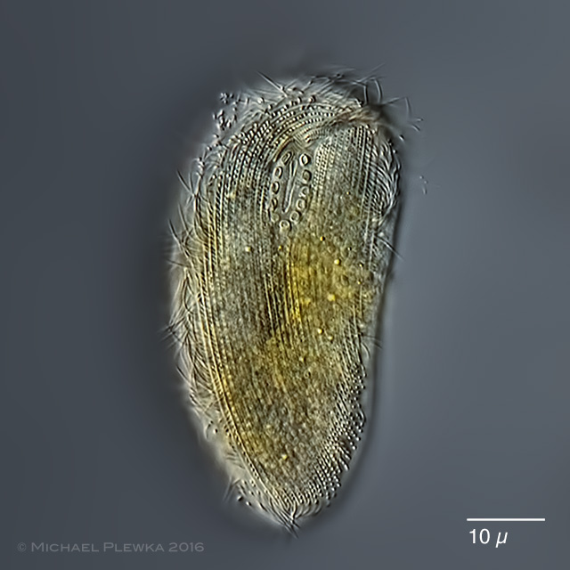
| Chlamydodon triquetrus: ventral view. Focus plane on the oval cytostome with nematodesmal rods. |
| |
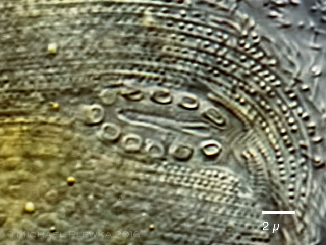 |
| Chlamydodon triquetrus: crop of the above image (turned 90 degrees clockwise). |
| |
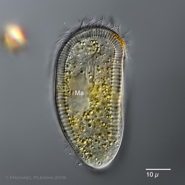 |
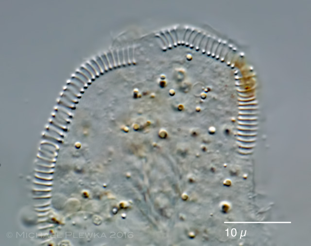
| Chlamydodon triquetrus: Focus plane on the macronucleus and the cross-striated band that surrounds the whole cell except for a small area at the posterior end (see next image). |
| |
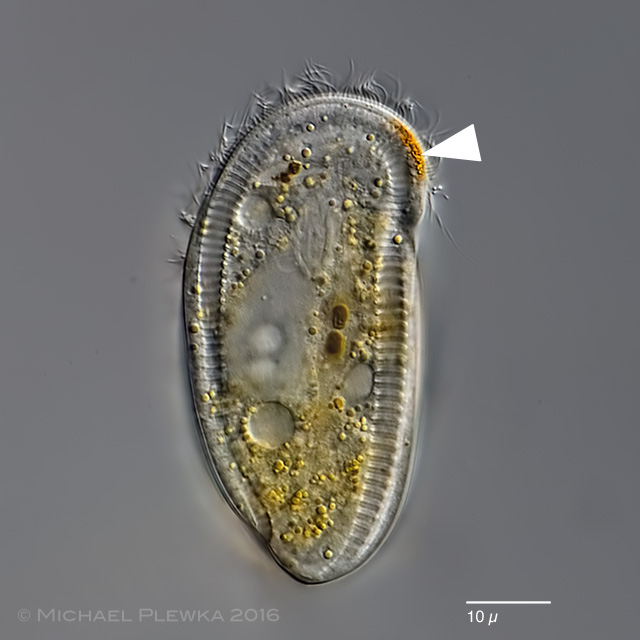 |
| Chlamydodon triquetrus: the cytoplasma contains many yellow granules that give the whole ciliate a yellowish color. Also visible are some contractile vacuoles and an aggregation of orange granules (arrowhead) that is also mentioned by KAHL (1935). |
| |
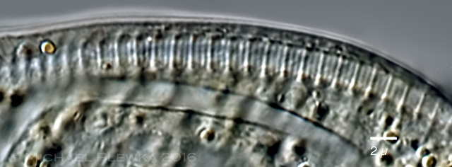 |
| Chlamydodon triquetrus: closeup of the cross-striated band |
| |
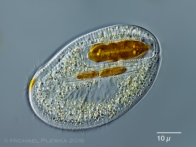 |
| Chlamydodon triquetrus: another specimen with two ingested diatoms. |
| |
 |
| Chlamydodon triquetrus: cross-striated band after treatment with destilled water. |
| |
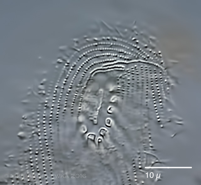 |
| Chlamydodon triquetrus: after treatment with destilled water also the pattern of the kinetids becomes visible. |
| |
| Location: Bad Frankenhausen; Kyffhäuser, Elisabethquelle |
| Habitat: detritus in salinic water (salinity 4-7%) |
| Date: 21.10.2016 |